Tonometry
Introduction: Measurement of Intraocular Pressure
- In veterinary patients, the single most consistent feature of all glaucomas characterized to date is elevation in intraocular pressure (IOP).
- Intraocular pressures > 25mmHg in dogs and > 31mmHg in cats, or differences of more than a few mmHg between eyes should be considered suspicious.
- However, many factors can influence IOP measurements,including breed, age and gender, method of restraint, time of day, the technique of the “tonometrist” and the tonometer itself. Hence, it is very difficult to provide a precise “cut-off” point for determining normal versus abnormal IOP. For this reason, one should always interpret tonometry readings in light of other clinical findings. “High” IOP is a measurement, not a diagnosis.
- Direct measurement – manometry
- The most accurate way to measure IOP
- Invasive, thus not practical in a clinical setting
- Restricted to a research setting
- Used in the validation of accuracy and precision of indirect tonometry methods
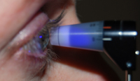
- Indirect Measurement – tonometry
- Two main forms that both represent contact tonometry:
- Indentation tonometry
- Applanation tonometry
- Non-contact (air puff) tonometry
A detailed review of the history of tonometry is out-with the scope of this lecture and the list of references provided is neither intended to be exhaustive, nor exhausting. For a detailed review of the history of tonometry in humans see Kniestedt, et al.[1] Chihara,[2] considers theoretical challenges to accurate determination of IOP in humans.
All methods of tonometry are subject to the same basic tenets. Extraneous factors that influence IOP should be avoided / taken into consideration:
- Pressure exerted by the fingers of the examiner on the globe or eyelids will artificially raise IOP.[3]
- Squeezing of the eyelids, eye movements and accommodation can falsely alter IOP
- Firm restraint, particularly where pressure is exerted on the neck (jugular occlusion) and tight collars increase episcleral venous pressure and elevate IOP. [3, 4]
- Posture / head position can influence IOP.[5, 6]
- Drugs administered, including sedation, may affect IOP.[7, 8]
- Repeated measurements over time (particularly where a weight is applied to the eye) will lower IOP by influencing pressure dependent aqueous outflow facility.
- Time of day influences IOP.
- All tonometers will show inter-operator variability to a greater or lesser extent.
- Different tonometers differ in their accuracy and precision.
- Age has an effect on IOP that is species dependent; with a decline in IOP noted in older dogs and cats.[9, 10]
- When evaluating studies that claim to validate a tonometric method in a given species:
- Consider whether values obtained are compared to manometry (the gold standard) or just another inherently inaccurate device…
- Consider accuracy (which is reflected by the slope of the regression line, this should be as close to one as possible)
- Consider reproducibility /consistency / variability (which is at least partly represented by the r2 value, which should also be close to 1).
- Any relative under- or over-estimation of IOP should preferably be linear and consistent over the range of IOPs that might be expected in clinical patients.
- Are all of the data points presented, to give the reader a better sense of the performance of the instrument?
- What is the magnitude of any departure from true IOP? 2mmHg may be of little clinical importance when distinguishing between a normal and glaucomatous subject, but may be of greater importance if the tonometer is to be used to detect a small change in IOP in a pharmacologic study.